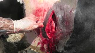
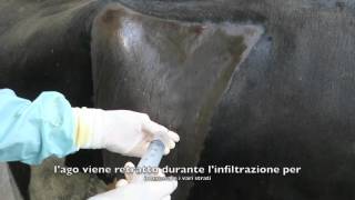
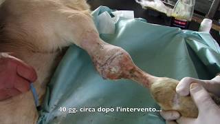
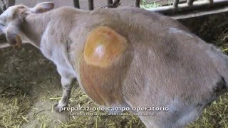
Jersey cow undergoing right-flank surgery for left displaced abomasum. During manual maneuvers to return the abomasum to its natural position, recent adhesions with abundant fibrin are found between the visceral peritoneum of the abomasum and the parietal peritoneum of the abdominal cavity. Proceeding cautiously, the adhesions are manually released via blunt dissection, then attempting to bring the abomasum back to the level of the laparotomy incision. If necessary, the incision is extended ventrally. In this case, a large amount of recently formed fibrin can be seen, which allowed us to completely free the abomasal wall from the abdominal wall. If adhesions are already fibrotic and impossible to resolve via blunt dissection, it may be useful to perform a left-flank laparotomy to address them directly.
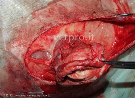
INFEZIONI OMBELICALI
Incisione ellittica cute attorno all'ombelico; apertura cavità addominale con incisione sulla lina mediana cranialmente ombelico.
View23 April 2026
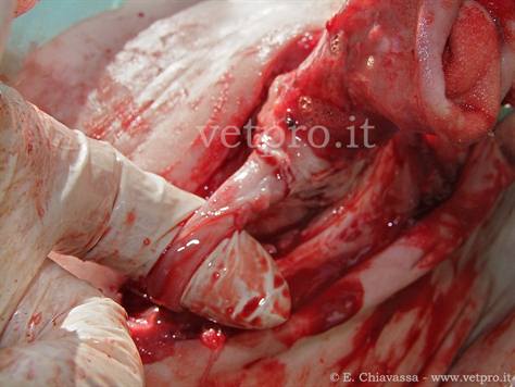
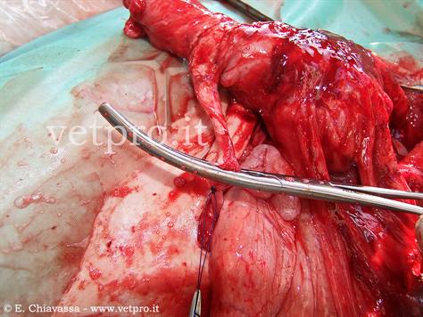
INFEZIONI OMBELICALI
Dopo incisione e disimpegno del pacchetto ombelicale dalla parete addominale, legatura (monofilamento assorbibile) e scontinuazione vena ombelicale.
View23 April 2026
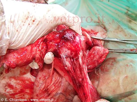
INFEZIONI OMBELICALI
Aderenza del grande omento al tratto addominale dell'uraco; risoluzione dell'aderenza per trazione.
View23 April 2026